
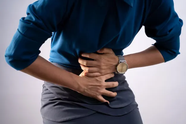

Renal calculi (kidney stones) are hard, crystal-like deposits that form in the kidneys when certain minerals become concentrated in urine. They can be tiny and pass unnoticed, or they can move into the ureter, the tube between kidney and bladder, and cause intense, wave-like pain. If you have received an ultrasound or CT report mentioning renal calculi, the most useful next step is not to panic over the term. Understand the stone’s size, location, symptoms, and whether it is affecting urine flow, then get the right medical advice.
Renal calculi definition: what does it mean?
The renal calculi definition is simple: they are solid collections of minerals and salts that form inside one or both kidneys. They are more commonly called kidney stones, renal stones, or urinary stones. In clinical language, renal calculi are also known as nephrolithiasis when referring to the condition of having kidney stones.
Urine normally carries dissolved minerals away from the body. When urine is too concentrated, or when the balance of substances that promote and prevent crystals changes, tiny crystals can stick together and grow. Stone formation is not always caused by one “wrong” food or habit. Low fluid intake, a previous stone, family history, certain bowel or metabolic conditions, some medicines, infection, and the type of stone can all matter.
The most common stone types include calcium oxalate, calcium phosphate, uric acid, struvite (often linked with infection), and cystine stones. Knowing the type, when a passed or removed stone can be analysed, helps make prevention more specific.
What symptoms can renal calculi cause?
A stone sitting quietly in the kidney may not cause symptoms. Trouble often begins when it moves or interferes with urine flow. Possible symptoms include:
- sharp or cramping pain in the side or back, often below the ribs;
- pain that comes in waves and may change in intensity;
- pain or burning while urinating;
- pink, red, or brown urine, which can indicate blood in the urine;
- nausea or vomiting;
- an urgent need to urinate, frequent small amounts of urine, or cloudy or foul-smelling urine; and
- fever or chills, especially when infection is present.
Symptoms can overlap with urinary tract infection, bowel problems, or other causes of abdominal pain. Do not assume that every backache or burning sensation is a kidney stone. For a related overview, you can learn about urinary tract infection symptoms, but fever, chills, severe pain, or urinary symptoms with a known stone need prompt medical advice.
Book a Confidential Consultation
Renal calculi referred pain: why does pain travel to the groin?
Renal calculi referred pain describes pain felt away from the kidney itself. A stone moving through the ureter can irritate tissues and trigger nerves that share pathways with the lower abdomen, groin, bladder, or genital area. This is why renal colic may start in the flank or back and travel down towards the lower abdomen or groin.
The location of pain can change as the stone moves, but pain location alone cannot tell you exactly where a stone is or how serious it is. Imaging and a clinician’s assessment are needed to check the stone’s size, location, and whether the kidney is obstructed.
Renal calculi 7 mm: is a 7 mm kidney stone serious?
Seeing renal calculi 7 mm on a scan report understandably causes concern. A 7 mm stone is not automatically an emergency, but it is large enough that you should not decide on home management based only on its size. Whether it may pass, cause problems, or need a procedure depends on where it is located, whether it is blocking urine flow, your level of pain, kidney function, and whether infection is present.
Small stones may sometimes pass with observation and prescribed pain management. Larger stones, stones that block the urinary tract, or stones causing severe pain, persistent vomiting, infection, or reduced kidney function may require more urgent urological treatment. A urologist may discuss options such as shock-wave lithotripsy, ureteroscopy, or another procedure, depending on the case.
Ask the clinician reviewing your scan: Is the 7 mm stone in the kidney or ureter? Is there swelling of the kidney or blockage? Is there evidence of infection? What symptoms mean I should go to emergency care? These questions are more useful than a generic “Will it pass?” answer online.
How renal calculi are diagnosed
Diagnosis usually combines your symptoms and medical history with urine tests, blood tests, and imaging. An ultrasound can identify many stones and check for swelling. In some situations, CT imaging gives more detailed information about stone size and location. A urine test may look for blood, infection, or crystals.
If you pass a stone, your clinician may ask you to keep it for laboratory analysis. For recurrent stones, a clinician may recommend a 24-hour urine collection to measure urine volume and mineral levels. This information makes diet and prevention advice more personal and meaningful.
Renal calculi diet chart: a practical guide, not a one-size-fits-all prescription
There is no single renal calculi diet chart that suits every person. The best dietary advice depends on the stone type, urine findings, medical conditions, and any medicines you take. For example, a person with calcium oxalate stones may need a different emphasis than someone with uric acid or cystine stones.
The guide below is a sensible starting point for many adults, but it should be personalised by a doctor or dietitian, especially if you have kidney disease, diabetes, heart failure, are pregnant, or take regular medicines.
Avoid starting stone-prevention supplements, high-dose vitamin products, or restrictive diets without clinical guidance. “Natural” does not always mean safe for every stone type or medical condition.
Renal calculi prevention: practical steps after a stone
Renal calculi prevention begins with finding out why the stone developed. If this is your first small stone, the plan may focus on fluids and follow-up. If stones keep returning, a more detailed assessment, such as stone analysis, blood tests, and 24-hour urine testing, can identify correctable factors.
For many people, prevention includes drinking enough fluid, reducing excess sodium, maintaining an appropriate dietary calcium intake, moderating excess animal protein, and following stone-type-specific advice. Some people may need prescribed medicines, such as citrate or other treatments, based on the stone type and urine results. Do not take these medicines or change doses without a qualified clinician.
Make prevention easier by building small habits: carry a water bottle, drink water at meals and after activity, check food labels for sodium, and keep follow-up appointments. In India’s hot weather, dehydration can become an important trigger, especially during long outdoor work, travel, or exercise.
When should you seek urgent medical help?
Do not wait for a routine appointment if a stone is suspected and you have any of the following:
- fever, chills, confusion, or feeling severely unwell;
- severe or worsening pain that is not controlled;
- repeated vomiting or an inability to keep fluids down;
- very little urine, inability to pass urine, or new swelling;
- visible blood in urine with significant symptoms; or
- pregnancy, a single functioning kidney, known kidney disease, or a weakened immune system along with possible stone symptoms.
These symptoms can indicate infection, dehydration, or urinary blockage. They need urgent evaluation, often by a urologist or emergency service, rather than self-treatment.
Treatment: why the scan report and symptoms both matter
Treatment is chosen according to the stone’s size, location, composition, symptoms, and effect on the urinary tract. A clinician may monitor a small stone, provide medication for pain or nausea, advise on fluids when safe, or plan a procedure to break up or remove a stone. A urologist is the appropriate specialist when a stone is large, obstructing, infected, recurrent, or causing severe symptoms.
Supportive lifestyle care may help people improve hydration, diet consistency, and long-term habits. It must not delay urgent treatment or replace a urological assessment for a 7 mm stone, a blocked stone, or a possible infection. You can explore kidney stone consultations at Dr. Tathed’s Homeopathic Clinic for a diagnosis-aware discussion of symptoms and prevention; ensure that any suspected obstruction or infection is assessed promptly by an appropriate medical specialist.
Speak with Dr. Tathed’s Homeopathic Clinic
Kidney-stone symptoms and repeat stone episodes can be stressful. Dr. Tathed’s Homeopathic Clinic provides private consultations in PCMC-Pune (Chinchwad), Viman Nagar, Pune, and Thane West. Learn more about the clinic and care team, or book a confidential consultation at the location most convenient for you.
For safety, take your scan report and test results to the consultation. If you have severe pain, fever, chills, vomiting, or reduced urine output, seek urgent urological or emergency care first.
Find Your Nearest Homeopathy Clinic
- Homeopathy Clinic in Chinchwad (PCMC), Pune : Opposite lokmanya hospital, near railway bridge, Renuka Sai Society, Ganesh Nagar, Chinchwad, Pimpri-Chinchwad, Maharashtra 411033
- Homeopathy Clinic in Viman Nagar, Pune : Datta Mandir Chowk, Unit 106, 1st Floor, Lunkad Skymax Mall, Konark Nagar, Clover Park, Viman Nagar, Pune, Maharashtra 411014
- Homeopathy Clinic in Thane West : Office No. 207, JVM’s Corner Stone, Hariniwas Circle, Lal Bahadur Shastri Marg, Naupada, Thane West, Thane, Maharashtra 400602
Frequently asked questions about renal calculi
Renal calculi are also known as what?
Renal calculi are also known as kidney stones, renal stones, or urinary stones. The condition of having kidney stones is often called nephrolithiasis.
Is a 7 mm renal calculus dangerous?
A 7 mm stone requires timely clinical review because the right plan depends on its location, symptoms, urine flow, infection status, and kidney function. Seek urgent care if you have fever, severe pain, vomiting, reduced urine output, or feel very unwell.
Can a renal calculi diet chart dissolve a stone?
Diet can help lower the chance of future stones and support overall urinary health, but it is not a substitute for assessment or treatment of an existing stone. Diet plans should be adjusted to the stone type and your health needs.
Where is renal calculi referred pain felt?
Pain may start in the flank or back and travel towards the lower abdomen or groin. Because other conditions can cause similar pain, a clinician should confirm the cause.
How can I prevent renal calculi from coming back?
Drink enough fluids when medically safe, reduce excess sodium, follow stone-type-specific diet advice, and complete any recommended follow-up or urine testing. Your clinician may prescribe preventive medicine for some stone types.
Can I visit Dr. Tathed’s Homeopathic Clinic for kidney stone guidance?
Dr. Tathed’s Homeopathic Clinic offers consultations at PCMC-Pune (Chinchwad), Viman Nagar, Pune, and Thane West, as well as online consultations. You can book a confidential consultation to discuss your reports, symptoms, hydration, diet habits, and prevention questions. If you have a 7 mm stone, severe pain, fever, vomiting, or reduced urine output, do not delay urological or emergency assessment.
What is the difference between a kidney stone and a UTI?
A kidney stone is a solid mineral deposit; a UTI is an infection of the urinary tract. They can sometimes occur together and share symptoms such as burning urination or blood in the urine. Fever or chills with urinary symptoms needs prompt medical evaluation.
Medical references :
- NIDDK: Kidney stones - definition, symptoms, diagnosis and treatment
- NIDDK: Eating, diet and nutrition for kidney stones
- NIDDK: Treatment for kidney stones and prevention of recurrence
- Urology Care Foundation: Kidney stones - symptoms, diagnosis and treatment
Important medical note: This article is educational, not a diagnosis or a treatment plan. Sudden severe flank pain, fever or chills, vomiting, reduced urine output, or feeling very unwell can signal a stone with infection or blockage and needs urgent medical assessment.